What is persistent and recurrent thoracic outlet syndrome?
Persistent thoracic outlet syndrome (TOS) is a condition when the disease continues or even worsens after surgery. Recurrent thoracic outlet syndrome is slightly different from persistence. Recurrence means that symptoms of TOS return after a brief period of improvement. This short time frame is often called “honeymoon”. There are several reasons for failure.
Causes of Recurrent Thoracic Outlet Syndrome
Wrong diagnosis
There are many conditions that may mimic thoracic outlet syndrome. Cervical disc herniation, shoulder joint problems, fibromyalgia, and Raynaud’s disease may be misdiagnosed as TOS.
- Cervical disc herniation. There are many common features between TOS and disc herniation. Both diseases are associated with nerve compression. Consequently, cardinal symptoms like pain, numbness, and weakness are present in both diseases. Therefore, differential diagnosis can be challenging. To make things worse, some patients may simultaneously have cervical disc herniation and TOS.
- Fibromyalgia can be very easily mistaken for TOS and vice versa. The main symptom in fibromyalgia patients is pain. The painful area may be very similar to TOS, but numbness and weakness are uncommon in fibromyalgia.
- Raynaud’s disease is a chronic condition affecting arteries and leading to progressive narrowing. As a result, patients develop cold intolerance and ischemia in their arms and hands. Fingertips are particularly affected. The main difference between TOS and Raynaud’s phenomenon is the absence of other thoracic outlet-related symptoms like nerve injury and venous obstruction. Also, from a diagnostic standpoint, angiography in Raynaud’s disease shows progressive narrowing of all arm, hand, and finger arteries, while in arterial TOS, only the subclavian artery is compressed from outside above the first rib.
- Parsonage-Turner Syndrome is an extremely rare disorder affecting the brachial plexus. It has an acute onset, and the pain is excruciating. The pain is followed by severe weakness in the arm. Upon recovery, patients usually develop permanent paresis of the arm and hand muscles. The main differences between PTS and TOS are acute onset, severity, lack of pain after recovery, and absence of vascular involvement.
- Complex regional pain syndromes. There are two types of CRPS. Type 1 is previously known as reflex sympathetic dystrophy and is more frequent. Type 2 used to be called causalgia. These are highly debilitating syndromes significantly affecting patients’ quality of life. CRPS develop after relatively minor injury like trauma, fracture or surgery. With time the intensity of pain surpasses the initial injury and spreads to other areas. Usually hands, arms, feet and legs are affected. The degree of pain is disproportionate to original injury and there are additional symptoms. Autonomic nervous system is frequently involved (old name used to be reflex sympathetic dystrophy). There might be episodes of swelling, redness, coldness, sweating. Chronic inflammation is typical and there is also evidence of immune system involvement. The difference between CRPS types is the presence of peripheral nerve injury in type 2.
The diagnosis of thoracic outlet syndrome is made clinically, and radiographic confirmation is not always reliable. Misdiagnoses are very frequent and can produce poor outcomes. A crucial step towards success is a thorough evaluation conducted by an experienced specialist.
Poor surgical technique
Surgery is the most effective and long-lasting treatment for TOS. It aims for decompression of the neuro-vascular bundle and is called thoracic outlet decompression (TOD). However, insufficient surgical technique is by far the most common reason for recurrence. Post-operative failure can be attributed to two factors: TOD surgery without first rib resection and with incomplete rib resection.
Scalenectomy and neurolysis without rib resection
Historically, the first rib resection was the initial treatment for thoracic outlet syndrome. However, complications and technical difficulties associated with first and additional rib removal led to the proposal of an alternative first rib-sparing approach in the 1920s.1. The technique is mostly unchanged, and it is still used today. The anterior and middle scalene muscles are partially removed, and the neuro-vascular bundle is relieved from compressive soft tissue bands. This treatment gained a lot of popularity due to low complications, and technical simplicity. Subsequent research, however, demonstrated that it was unsuccessful for long-term control. Many surgeons began to voice concerns about the poor results of this approach in the 1940s, and by the 1950s and 1960s, it was clear that this treatment ought to be discontinued.2. 3. Results improved right away when surgeons switched back to the first rib removal procedure.
Furthermore, rib removal was shown to be better in a prospective randomized trial.4. It may appear that scalenectomy and neurolysis should have been abandoned long ago in the light of scientific and clinical evidence. Regretfully, a lot of surgeons continue to treat TOS with this technique today.
Incomplete first rib removal
Without doubt, first rib removal is an indispensable part of thoracic outlet decompression surgery. Yet, the degree of first rib resection varies significantly. Depending on the surgeon’s skills and technique, the extent of bone removal ranges from mere touching to near complete rib resection. There is strong scientific evidence indicating that the degree of first rib resection is the most powerful factor affecting long-term success.
Typically, TOD surgery is performed either from the front of the neck (supraclavicular approach) or from the armpit (transaxillary approach). Yet, it is technically difficult and risky to totally remove the first and accessory ribs via these approaches. The reason is the anatomical relation of the first rib. The first rib has joint connections with the spine posteriorly and with the sternum anteriorly. The posterior and anterior regions of the first rib are very deep and hard to access.
Anatomically the brachial plexus, subclavian vein and subclavian artery pass over the first rib. These are the delicate structures which should be protected at all cost. Yet in order to access the first rib these nerves and vessels should be moved to expose the bone. Like the first rib, the accessory cervical rib posteriorly has either a joint or bony connection with the spine. The proximity to the brachial plexus is even more for the accessory rib than for the first rib, making safe exposure and total removal nearly impossible. Therefore, surgeons prefer not to risk these frail, vital structures and deep section parts remain untouched.
On the other hand, the middle section of the first rib and the tip of the accessory rib are relatively easy and safe to access and respect. Thus, in the vast majority of cases, only the midst of the first is removed. The anterior and/or posterior thirds of the first rib are usually left untouched, along with the main portion of the accessory rib.
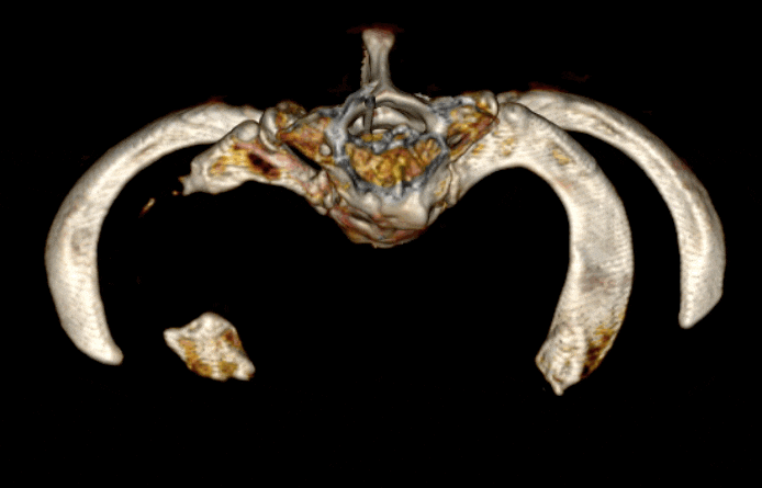
The remaining rib parts are the main cause of persistence or recurrence after surgery. It has been confirmed that the size of remaining bones correlates with the likelihood of recurrence.5.6. Almost a quarter of patients treated with conventional methods do not benefit from surgery, and another quarter have remaining symptoms.7. Many patients worsen after surgery. This phenomenon was explained by Dr Robert Leffert, who coined the term “missing third”.8. With the middle section of the first rib removed, the brachial plexus sticks to the posterior rib stump and the subclavian vein to the anterior stump. Normally, nerves and vessels do not adhere to the bone. However, after surgery, scar tissue develops in the surgical field and sticks everything indiscriminately. This scar tissue anchors nerves and vessels to the bone. The subclavian artery is usually sparred because there is no bone left underneath, and it usually sinks down with surrounding soft tissue. This soft tissue “sinking” pulls the nearby brachial plexus and subclavian vein down. With remaining rib stumps underneath and immobility due to scar tissue, both the brachial plexus and subclavian vein get strained, leading to worsening.
Treatment for persistent and recurrent thoracic outlet syndrome includes repeat surgery. However, redo surgery is more challenging due to distorted anatomy and scar tissue. Only experienced surgeons should attempt to undertake corrective procedures for previously unsuccessful TOS surgeries.
Next: PURED Procedure for Thoracic Outlet Syndrome
References
- Adson AW, Coffey JR. Cervical Rib: A Method of Anterior Approach for Relief of Symptoms by Division of the Scalenus Anticus. Ann Surg. 1927;85(6): 839-857. https://doi.org/10.1097/00000658-192785060-00005 [↩]
- Clagett OT. Research and prosearch. J Thorac Cardiovasc Surg. 1962;44: 153-66. https://pubmed.ncbi.nlm.nih.gov/13879636/ [↩]
- Raaf J. Surgery for cervical rib and scalenus anticus syndrome. J Am Med Assoc. 1955;157(3): 219-223. https://doi.org/10.1001/jama.1955.02950200017005 [↩]
- Sheth RN, Campbell JN. Surgical treatment of thoracic outlet syndrome: a randomized trial comparing two operations. J Neurosurg Spine. 2005;3(5): 355-363. https://doi.org/10.3171/spi.2005.3.5.0355 [↩]
- Likes K, Dapash T, Rochlin DH, Freischlag JA. Remaining or residual first ribs are the cause of recurrent thoracic outlet syndrome. Ann Vasc Surg. 2014;28(4): 939-945. https://doi.org/10.1016/j.avsg.2013.12.010 [↩]
- Mingoli A, Sapienza P, di Marzo L, Cavallaro A. Role of first rib stump length in recurrent neurogenic thoracic outlet syndrome. Am J Surg. 2005;190(1): 156. https://doi.org/10.1016/j.amjsurg.2004.11.006 [↩]
- Suzuki T, Kimura H, Matsumura N, Iwamoto T. Surgical Approaches for Thoracic Outlet Syndrome: A Review of the Literature. J Hand Surg Glob Online. 2023;5(4): 577-584. https://doi.org/10.1016/j.jhsg.2022.04.007 [↩]
- Leffert RD. Complications of surgery for thoracic outlet syndrome. Hand Clin. 2004;20(1): 91-98. https://doi.org/10.1016/s0749-0712(03)00084-2 [↩]